
Choosing a suture anchor is not a simple catalog decision. It affects fixation strength, tissue handling, surgical time, and rehabilitation planning. In Suture Anchor Orthopedics, small differences matter: thread design, eyelet shape, insertion method, and anchor material can change performance inside fragile bone.
Market research reflects this demand. Grand View Research identifies orthopedic devices as a steadily expanding global sector, supported by sports injuries, aging populations, and minimally invasive procedures. MarketsandMarkets also reports continued growth in orthopedic implants, including fixation technologies. These reports describe market direction, not a guarantee of clinical success. Product selection still requires evidence.
Dr. Freddie H. Fu, a respected sports medicine surgeon, often emphasized, “Anatomy is the key to successful surgery.” That principle belongs at the center of every anchor purchase. Buyers should examine whether the anchor matches the repair site, bone quality, suture configuration, and surgeon training. A lab-tested pullout value may not predict performance in osteoporotic or irregular bone.
Look closely.
Review peer-reviewed studies, independent testing, sterilization data, and manufacturer instructions. Confirm whether the device has appropriate regulatory clearance in your market. Ask how easily the anchor advances through dense cortical bone. Check the suture’s abrasion resistance after knot tying. These details become tangible in the operating room, where a damaged strand or poorly seated anchor can disrupt an otherwise careful repair.
No product is perfect. Comparative studies may use different models, loads, and endpoints. That limitation deserves attention. The following five tips offer a practical framework for comparing Suture Anchor Orthopedics products with greater discipline, transparency, and clinical judgment.
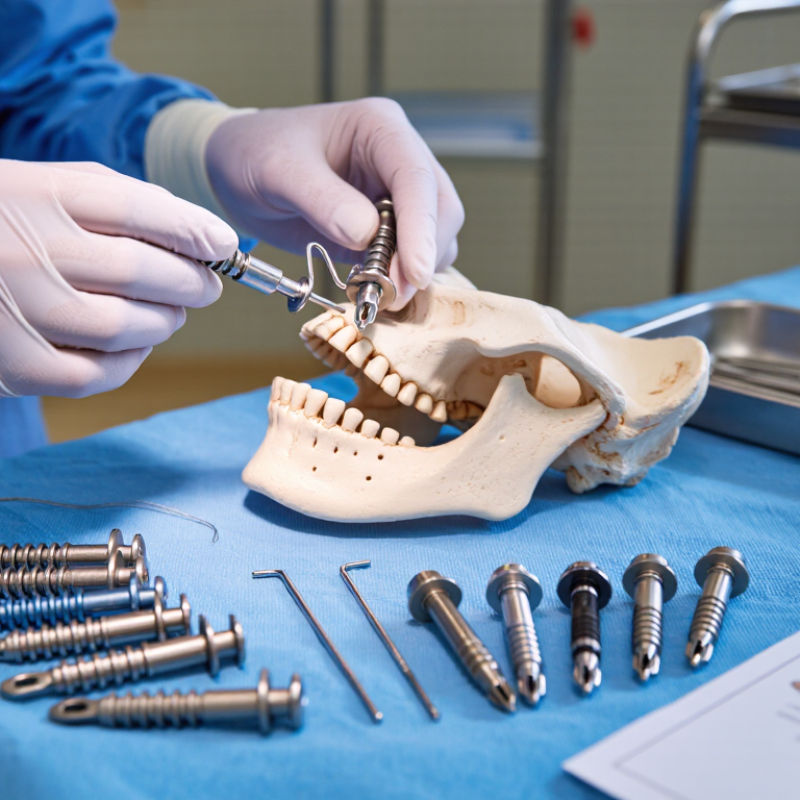
5 Tips for Buying Suture Anchors for Orthopedics
Define the surgical requirement before comparing prices or packaging. Identify the procedure, tissue quality, tear pattern, fixation site, and expected loading. A rotator cuff repair needs different planning from labral or foot-and-ankle fixation. Anchor diameter, insertion angle, thread configuration, and deployment depth must match the surgeon’s technique. Small details matter.
Clinical data should guide the specification. A 2023 review in Arthroscopy reported retear rates commonly ranging from 10% to 40% after rotator cuff repair, depending on tear size, tissue quality, and follow-up duration. This does not mean the anchor caused failure. Patient biology and repair technique also matter. The finding still supports careful selection of fixation strength, suture handling, and backup options. I have seen teams overbuying anchors without defining these variables. That habit deserves scrutiny.
Review material behavior, pullout resistance, knot security, imaging compatibility, and sterilization records. Ask for published test methods, not only headline strength values. The FDA’s MAUDE database shows why post-market event monitoring deserves attention, although reports do not prove causation. Check lot traceability, shelf life, instrument compatibility, and staff training before approval. Keep the evaluation clinical. A low unit price can become expensive when insertion is difficult or revision risk rises. The best purchasing file links each anchor choice to a specific surgical requirement, supported by evidence and surgeon feedback.
| Tip | Surgical Requirement | Typical Orthopedic Application | Suitable Anchor Configuration | Material Considerations | Suture Requirements | Key Specification to Check | Buying Checklist |
|---|---|---|---|---|---|---|---|
| 1 | Define the tissue and repair site before selecting the anchor. | Rotator cuff, labral, meniscal, ligament, tendon, or osteochondral repair. | Small-diameter anchor for limited bone stock; larger or multi-anchor constructs for higher-load repairs where clinically appropriate. | Metallic, polymeric, bioabsorbable, or biocomposite options may be available. Selection should match bone quality, healing objectives, and the device instructions for use. | Confirm the number of suture limbs, suture size, suture type, and whether the construct supports simple, mattress, or rip-stop configurations. | Anchor diameter, insertion depth, thread design, pullout or fixation data, and compatibility with the intended repair technique. | Match the anchor to the anatomical site, bone density, repair pattern, and surgeon-defined fixation plan. |
| 2 | Match the fixation method to the surgical approach and available working space. | Arthroscopic shoulder, hip, knee, ankle, hand, foot, or open soft-tissue procedures. | Preloaded anchors can reduce handling steps; knotless anchors can simplify tensioning; knotted anchors may provide suture-tension flexibility. | Select radiopaque or radiolucent characteristics according to postoperative imaging needs and the relevant device documentation. | Verify suture management features, loading sequence, eyelet or channel design, and whether the sutures are color-coded for handling. | Delivery-system diameter, insertion angle, cannula compatibility, drill or punch requirements, and access limitations. | Confirm that the delivery instrument fits the portal, incision, guide, and planned trajectory. |
| 3 | Assess bone quality and the expected mechanical demand of the repair. | Osteoporotic bone, revision surgery, high-tension tendon repair, or ligament reconstruction. | Consider screw-in or other insertion designs that provide appropriate engagement for the documented bone conditions and surgical technique. | Material stiffness, thread geometry, insertion torque, and potential degradation profile should be reviewed together rather than in isolation. | Use the suture size and construction supported by the anchor's validated mechanical data; avoid assuming that a larger suture always improves fixation. | Indicated bone quality, insertion torque range where provided, cyclic loading results, pullout data, and recommended drilling protocol. | Review mechanical testing conditions and confirm that they are relevant to the planned anatomical site and repair construct. |
| 4 | Consider imaging, revision, and long-term follow-up requirements. | Patients likely to require MRI follow-up, revision surgery, or evaluation of postoperative bone and soft tissue. | Select the anchor type according to its imaging labeling, removability, visibility, and intended permanence. | Metallic anchors may create susceptibility artifact; polymeric and bioabsorbable materials may offer different imaging characteristics. Always verify the specific MR labeling. | Confirm whether the suture is intended to remain permanently and whether it has features that support later identification or retrieval. | MR safety status, conditional scanning limits, artifact information, radiographic visibility, and revision procedure guidance. | Obtain the current MR conditions and implant identification information before purchase and clinical use. |
| 5 | Verify regulatory, sterilization, and supply requirements for the intended use. | Routine primary repair, urgent cases, ambulatory surgery, hospital procurement, or multi-site inventory management. | Sterile, single-use anchors with procedure-specific instruments and clearly defined indications. | Check biocompatibility information, sterilization method, shelf life, packaging integrity, and storage conditions. | Confirm the supplied suture is sterile, securely attached, correctly labeled, and included in the approved device configuration. | Intended-use statement, anatomical indications, contraindications, lot traceability, expiration date, and instrument availability. | Purchase only products cleared or approved for the relevant market and use them according to the current instructions for use. |
Note: Product specifications, indications, mechanical performance, MR labeling, and surgical technique vary by device. Confirm all purchasing and clinical decisions against the current instructions for use and applicable regulatory requirements.